Lung Cancer Detection and Diagnosis
Multiple tests are used to detect, diagnose, and learn the extent of a lung cancer diagnosis. The goal of diagnostic testing is to determine the exact size and location of the tumor(s) and the type of lung cancer. The tests used for each patient will vary based on the symptoms that are present. For those with a history of smoking, screening tests may be available before symptoms appear to find it as early as possible – when it’s easier to treat.
The cancer care team will also need to know if the cancer is affecting lymph nodes or other organs. This may be included in the diagnostic testing or it could be done after a diagnosis is determined.
Detecting Lung Cancer
One of the first steps in testing for lung cancer is to run imaging tests. These tests involve using X-rays, magnetic fields, or radioactive substances to create images of the inside of the body. The images will indicate if there is a tumor in the lungs and possibly other areas of the body. The most common imaging tests used to detect lung cancer include:
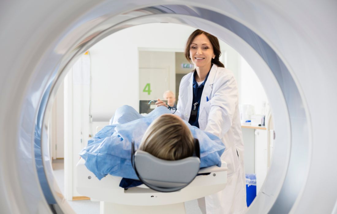
CT scan of the brain, chest, and abdomen: A series of detailed pictures of areas inside the body, taken from different angles. The images are made by a computer linked to an x-ray machine. A dye may be injected into a vein or swallowed to help the organs or tissues show more clearly. This procedure is also called computed tomography, computerized tomography, or computerized axial tomography.
PET scan (positron emission tomography scan): A small amount of radioactive glucose (sugar) is injected into a vein, which aims to find malignant tumor cells in the body. The PET scanner rotates around the body and makes a picture of where glucose is being used in the body. Malignant tumor cells show up brighter in the picture because they are more active and take up more glucose than normal cells.
MRI scan: This type of imaging allows the oncology team to identify areas where the lung cancer may have spread or to measure a tumor’s size. Because MRIs don’t work as well for body parts that are moving, like the lungs, they’re not normally used as a primary imaging tool for lung cancer diagnosis unless metastasis is suspected.
Bone scan: This test is also known as a radionuclide bone scan. It is used to check if there are cancer cells in the bones. A minimal amount of radioactive material is injected into a vein and travels through the bloodstream. The radioactive material collects in the bones and is detected by a scanner.
Identifying Lung Cancer Early
Some patients are diagnosed with lung cancer through screening. Although screening is not available to everyone, people with a history of smoking should get with their primary care physician to see if you're eligible.
Newly DiagnosedIndividuals at high risk for lung cancer should undergo annual lung screenings using a low-dose CT scan (LDCT), particularly because lung cancer symptoms often do not show up until later stages.
Lung cancer screenings are recommended for those who meet all of the following criteria:
Are between 50 and 80 years old and in good health, and
Currently smoke or have quit within the past 15 years, and
Have at least a 20-pack-year smoking history. (This is calculated by multiplying the number of packs of cigarettes smoked per day by the number of years smoked. For example, if someone smoked 2 packs a day for 10 years [2 x 10 = 20], they have 20 pack-years of smoking. The same applies to someone who smoked 1 pack a day for 20 years [1 x 20 = 20].)
Confirming a Lung Cancer Diagnosis
If imaging tests show signs of a tumor in the lungs, a definitive diagnosis is made by examining lung cells in the laboratory. This means a biopsy is needed. If cancer is confirmed, the biopsy report will also indicate the type of lung cancer that’s present.
Lung Biopsy / Tissue Sampling
Most lung cancer patients will have a biopsy of the lungs. This procedure collects a sample of lung tissue to be tested in a lab for cancerous cells. Various procedures can be used. Your oncologist and cancer surgeon will discuss what is best in your situation. Some of the types of tissue tests that may be performed include:
Fine-needle aspiration (FNA) biopsy of the lung: A CT scan, ultrasound, or other imaging procedure is used to locate the abnormal tissue or fluid in the lung, and then a small incision may be made in the skin where the biopsy needle is inserted into the abnormal tissue or fluid. A sample is removed with the needle and sent to the laboratory. A pathologist then views the sample under a microscope to look for cancer cells. A chest x-ray is performed afterward to ensure no air leaks from the lung into the chest.
Bronchoscopy: Uses a bronchoscope, which is a thin, tube-like instrument with a light and a lens for viewing, that is inserted through the nose or mouth into the trachea and lungs to look inside the trachea and large airways in the lung for abnormal areas. A bronchoscope may also have a tool to remove tissue samples, which are checked under a microscope for signs of cancer.
If a biopsy is not possible, your doctor may use other tests to confirm a diagnosis.
Other Lung Cancer Diagnostic Tests
In addition to a physical examination and discussion about your family health history, the following tests may be used to diagnose and stage both small cell lung cancer (SCLC) and non-small cell lung cancer (NSCLC):
Sputum Cytology: A microscope is used to check for cancer cells in the sputum (mucus) coughed up from the lungs.
Thoracoscopy: A surgical procedure to check for abnormal areas by looking at the organs inside the chest. A thoracoscope is a thin, tube-like instrument with a light and a lens for viewing. Typically, an incision (cut) is made between two ribs to insert a thoracoscope into the chest for viewing or for using a tool to remove tissue or lymph node samples that are then checked under a microscope for signs of cancer.
Thoracentesis: A needle is used to remove fluid from the space between the lining of the chest and the lung. A pathologist then views the fluid under a microscope to look for cancer cells.
Mediastinoscopy: A procedure in which a sample of the lymph nodes in the center of the chest underneath the breastbone is taken by making a small incision at the top of the breastbone. This procedure also requires general anesthesia and is done in an operating room.
Thoracotomy: A procedure in which the surgeon makes an incision in the chest to examine the lung directly and take tissue samples for testing. A thoracotomy is the procedure surgeons most often use to remove a lung tumor completely.
Laboratory tests: This involves testing samples of tissue, blood, urine, or other substances in the body. These tests help to diagnose disease, plan and check treatment, or monitor the disease over time.
Lung Cancer Treatment in Brevard County, Florida
At Cancer Care Centers of Brevard, our patients receive personalized cancer care and the latest treatments for lung cancer. If you were recently diagnosed with lung cancer, request an appointment with one of our lung cancer doctors today for a consultation and to discuss which treatments are available for you. Our cancer centers are conveniently located in Melbourne, Merritt Island, Rockledge, and Palm Bay, Florida, so you can receive quality cancer care close to home.