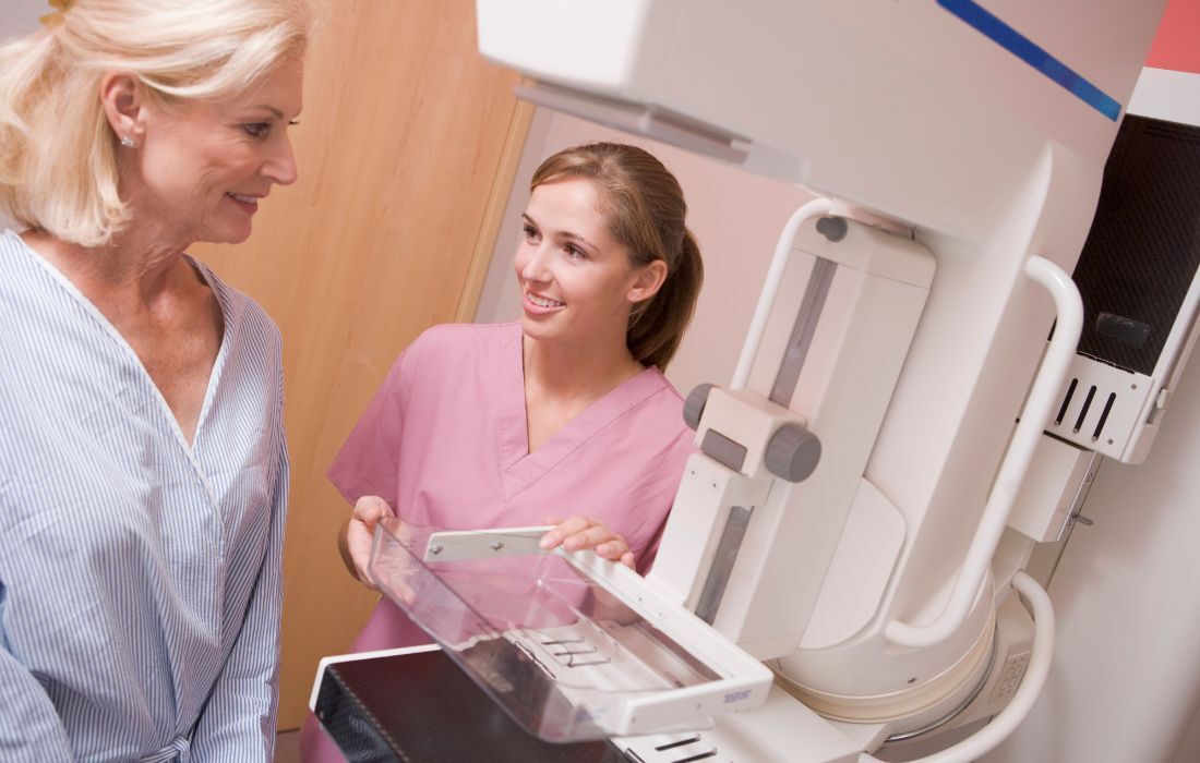
Breast Cancer Detection & Diagnosis
Even though breast cancer affects far too many women, the good news is that it can be detected early through a mammogram. Often, women experience little to no symptoms of breast cancer, making regular screenings especially important. The earlier breast cancer is detected, the easier it is to treat successfully.
If your routine mammogram comes back with abnormal results, your doctor may recommend a diagnostic mammogram and possibly a breast ultrasound for further investigation. Abnormal results do not necessarily mean that cancer is present; it simply means additional testing is needed to find out.
Screening Mammogram
A mammogram is an X-ray picture of breast tissue that can identify a breast lump before it can be felt or a cluster of calcium specks, called microcalcifications. Lumps or specks can be from cancer, precancerous cells, or other conditions. Further testing is needed to determine whether abnormal cells are present.
While 2D mammography has traditionally been the standard for breast cancer screening, that is no longer the case. Newer technology, known as 3D mammography or tomosynthesis, is now available. Unlike the single, straightforward X-ray image produced by 2D mammography, 3D mammograms capture images from multiple angles, providing a more comprehensive and detailed view of breast tissue.
All women benefit from 3D mammography, but especially those with dense breasts. Not all breast imaging centers offer 3D mammograms, so it's important to ask about this option when scheduling a screening appointment.
The American Cancer Society's screening recommendations differ based on whether a woman is at average or high risk for breast cancer.
Who Is Considered High Risk for Developing Breast Cancer?
Women who meet any of the following criteria are considered to have a higher risk of developing breast cancer:
A first-degree relative (mother, sister, or daughter) who has had breast cancer
Dense breast tissue
A known inherited genetic mutation, such as BRCA1 or BRCA2
Multiple family members on either side of the family who have had breast or ovarian cancer
Exposure to chest radiation before age 30
Other high-risk situations, as determined by your healthcare provider
Your doctor may suggest assessing your lifetime risk by completing a breast cancer risk questionnaire. For those with lifetime risks of 20% or higher, it is recommended to follow a high-risk screening process, which may include MRI surveillance and annual mammograms at an earlier age than average-risk women.
Discussing breast cancer risk with your doctor can help determine the right time to start breast cancer screenings.
Breast Screening Guidelines for Average Risk Women
Because breast cancer doesn’t show symptoms until the cancer has been present for a while, it’s important to use mammograms to detect breast cancer early. According to the American Cancer Society:
Women between the ages of 40 and 44 have the choice to start yearly mammography
Women aged 45 to 54 are recommended to receive a mammogram every year
Women age 55 and older can switch to having a mammogram every two years or continue yearly screening if they choose
Women under the age of 40 who have risk factors for breast cancer should ask their health care provider whether to have mammograms and how often to have them.
Some doctors will also recommend a baseline mammogram at age 35 so they can compare future mammograms to one when you were younger.
Diagnostic Mammogram
If the mammogram shows an abnormal area within the breast, your doctor may order clearer, more detailed images of that area. Doctors use diagnostic mammograms to learn more about unusual breast changes, such as a lump, pain, thickening, nipple discharge, or a change in breast size or shape. Diagnostic mammograms may focus on a specific area of the breast, offering more detailed views than traditional screening mammograms.
An Abnormal Area Has Been Found: What Now?
Other imaging tests may be ordered if an abnormal area is found on your mammogram. These can include:
Breast Ultrasound: An ultrasound uses sound waves to create a picture of the breast tissue. The sound waves echo differently when bouncing off abnormal tissue and healthy tissue. An ultrasound can distinguish between a solid mass, which may be cancer, and a fluid-filled cyst, which is usually not cancer.
Breast MRI: An MRI uses magnetic fields to produce detailed images of the body. They are used to check for additional disease or to determine how much the cancer has grown or spread. MRI is mainly reserved for women who are considered to be at a higher risk of developing breast cancer.

If Imaging Indicates Breast Cancer, Breast Biopsy is Next
If an abnormal area is found during a mammogram and other imaging tests, you will likely need a biopsy. A biopsy is the removal of tissue to look for cancer cells. It is the only way to tell for sure if cancer is present.
Types of Breast Biopsies
Your doctor may refer you to a surgeon or breast cancer specialist for a biopsy. The surgeon or doctor will remove fluid or tissue from your breast in one of several ways, with the core biopsy being the most common type.
Fine-needle aspiration biopsy: Typically used when a suspicious lump is filled with fluid. It involves using a thin needle to remove cells or fluid from a breast lump. An ultrasound may be used to help guide the needle.
Core biopsy: For this type of biopsy, the surgeon uses a wide, hollow needle to remove a small sample of suspicious breast tissue. Using imaging technology as a guide for the biopsy, a small marker may be inserted in the breast to mark the exact location of the biopsy. This makes it easier to find the tumor later during surgery. If no additional treatment is required after the biopsy, this marker can be seen in future mammograms to note where a biopsy was previously done.
Excisional biopsy: This type of biopsy removes the entire abnormal area with some margin of normal tissue. A marker is often placed before the procedure to ensure the surgeon goes directly to the correct location. Excisional biopsies are usually done while under anesthesia. This is usually done if the core biopsy didn’t provide conclusive results.
Skin biopsy: Typically used when there is a red or abnormal area on the skin of the breast that is not a common breast infection or allergic reaction. It involves using a cookie-cutter-like device to remove a small skin sample with a small amount of surrounding tissue.
After the biopsy, a pathologist will check the tissue or fluid removed from your breast for cancer cells.
If cancer cells are not detected, this means the lump is benign or non-cancerous.
If cancer cells are detected, the report will provide information to determine breast cancer type. The most common type of breast cancer that is diagnosed is invasive ductal carcinoma.
Lab Tests with Breast Tissue to Determine Treatment
Once breast cancer has been confirmed through diagnosis, specific lab tests are typically performed to determine the most effective treatment approaches. These tests may include:
Hormone receptor tests: Some breast cancers are fueled by hormones and may have receptors for estrogen, progesterone, or both. If these tests indicate the presence of hormone receptors, hormone therapy is often recommended as a treatment option.
HER2/neu test: The HER2/neu protein is found on certain cancer cells. This test determines whether there is an overabundance of HER2/neu protein or an excess of its gene copies. Tumors with increased levels of HER2/neu often benefit from targeted therapy as part of their treatment plan.
Learn more about how hormone receptor status plays a role in breast cancer.
It may take several days to receive these test results. These results are essential for guiding your oncologist in selecting the most appropriate breast cancer treatment options for you.